Cranial tibial muscle
Musculus tibialis cranialis
Flexes the hock.
- System
- Muscle
- Region
- Pelvic limb
- Sides
- Paired: left and right
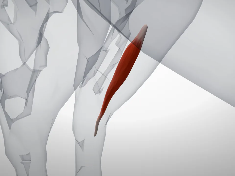
Where it runs from and to
A muscle is told apart from its neighbours by what it takes hold of at each end, which is also what decides what it does when it shortens.
- Origin
- The proximal end of the tibia, on its lateral surface.
- Insertion
- The third metatarsal and the tarsal bones.
- Action
- Flexes the hock.
- Nerve
- The deep fibular (peroneal) nerve.
Written for Vetomica in its own words from the standard reference anatomy of the horse; the books it was checked against are listed below.
Terminology
- English
- Cranial tibial muscle
- Latin (NAV)
- Musculus tibialis cranialis
- Status
- A valid term in the Nomina Anatomica Veterinaria
Read next
- Biceps muscle of the thighSame region
- Calcaneus (fibular tarsal bone)Same region
- Central tarsal boneSame region
- FemurSame region
- FibulaSame region
- Fourth tarsal boneSame region
- Gastrocnemius muscleSame region
- Os metatarsale IISame region
- Os metatarsale IVSame region
- PatellaSame region
- SemitendinosusSame region
- Semitendinous muscleSame region