Thorax
Pleural effusion
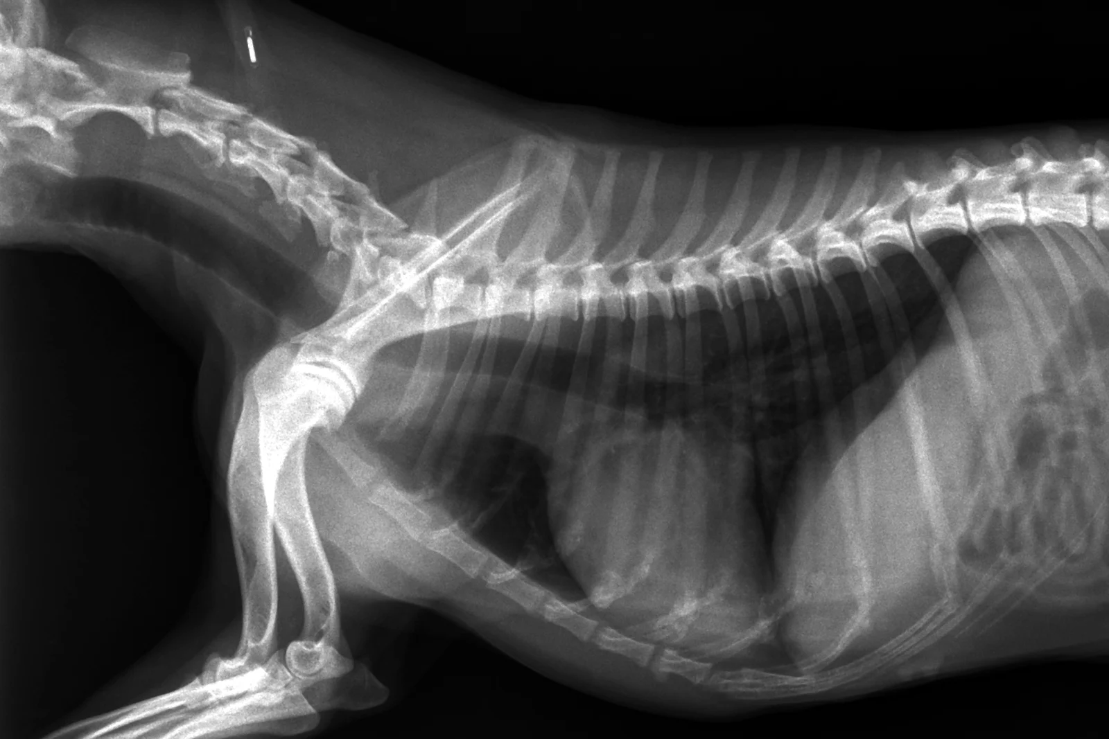
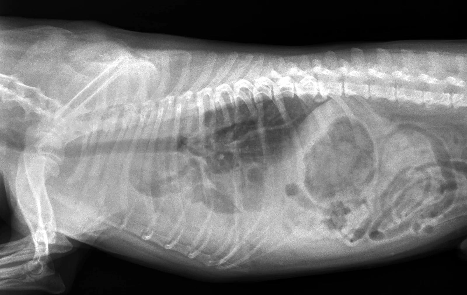
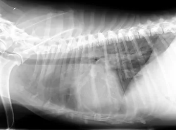
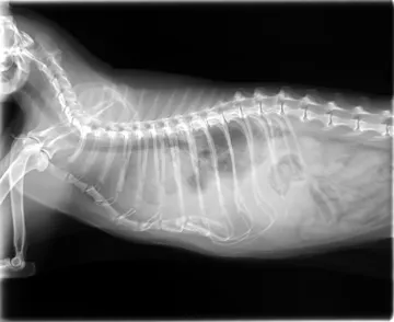
Fluid in the chest. Fluid in the pleural space, between the lungs and the chest wall. Fluid has the opacity of soft tissue, so it blurs and hides the edges of whatever it touches.
What to look for
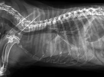
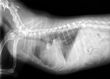
- Lung lobes pulled away from the chest wall, with a soft-tissue band between lung and ribs.
- Fissure lines: thin opaque lines between the lobes.
- The heart and diaphragm outlines blurred or lost (border effacement).
- Lobe edges rounded or scalloped rather than sharp.
Common causes
Heart failure, chylothorax, pyothorax, neoplasia, haemorrhage, feline infectious peritonitis in cats.
Against the normal
Normally the lungs reach the chest wall and the heart and diaphragm have crisp edges.
Radiographs: VetXRay, Banzato T., Burti S., Zotti A., Wodzinski M., University of Padua, CC BY 4.0. Converted to web images; findings as annotated by the dataset's radiologist. Dataset · Licence. For learning: not a diagnostic service.