Vetomica Imaging · Radiography and CT
Read a chest film. Normal first, then what changes.
Real dog and cat radiographs, each read by a radiologist. Start from a labelled normal and open any structure in 3D, then put each common finding beside it.
Normal canine thorax, lateral view
Left lateral, head to the left. Choose a number to name the structure and see it in the 3D dog.
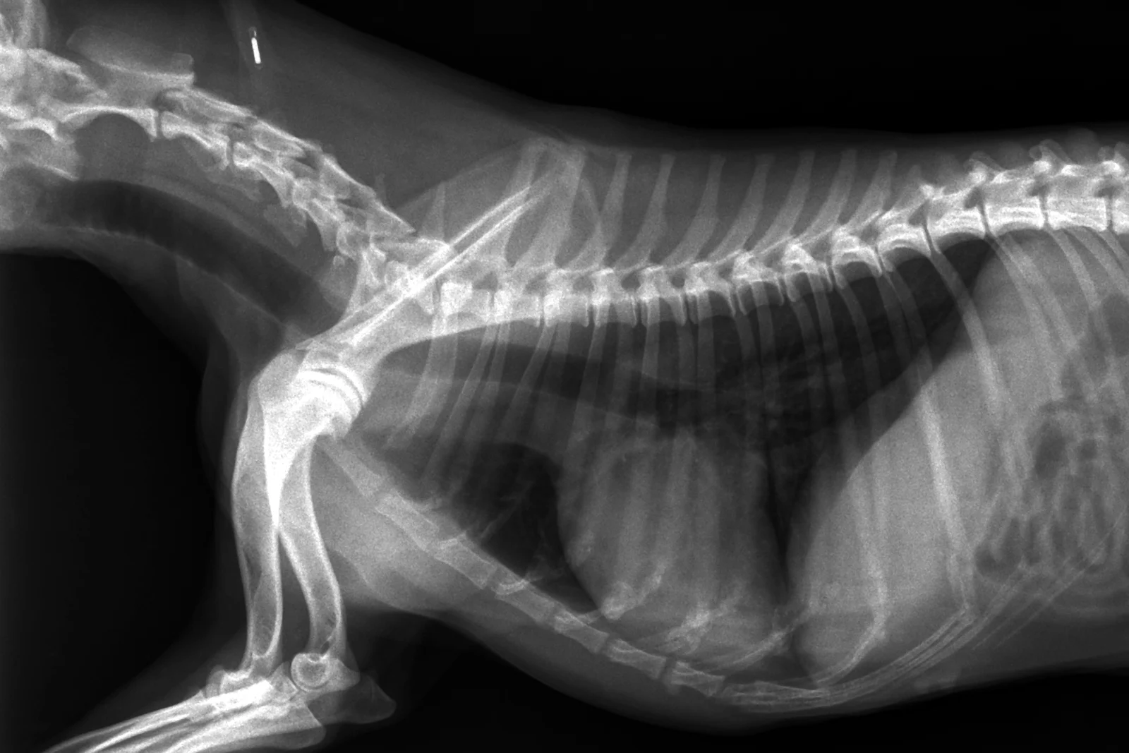
The cardiac silhouette: about two and a half to three and a half intercostal spaces wide on a lateral view.
See it in 3DFindings
Each one beside the normal film, with what to look for.
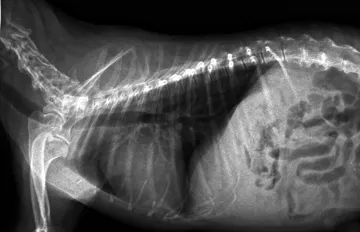 CardiomegalyAn enlarged heart
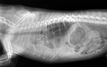 Pleural effusionFluid in the chest
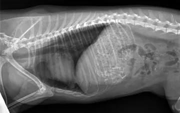 PneumothoraxAir around the lungs
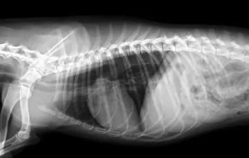 MegaoesophagusA dilated oesophagus
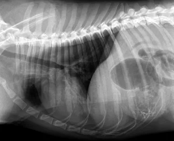 Alveolar patternAir spaces filled
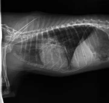 Bronchial patternThickened airway walls
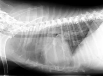 Interstitial patternA hazy lung
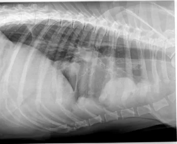 Thoracic massA mass in the chest
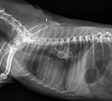 Foreign bodySomething that should not be there
CT of the head
Real CT scans you scroll in three planes: normal skulls of four head shapes and a cat, and a head-trauma case.
Open the CT scansMRI of the brain
Normal dog brains in fine detail, and a head-trauma case in T1, T2, FLAIR and contrast, plane by plane.
Open the MRI scansUltrasound
Kidney, bladder, liver, spleen, gut, adrenals and heart in dogs and cats: the normal image beside the common findings.
Open the ultrasound imagesRadiographs: VetXRay, Banzato T., Burti S., Zotti A., Wodzinski M., University of Padua, CC BY 4.0. Converted to web images; findings as annotated by the dataset's radiologist. Dataset · Licence. For learning: not a diagnostic service.