Thorax
Alveolar pattern
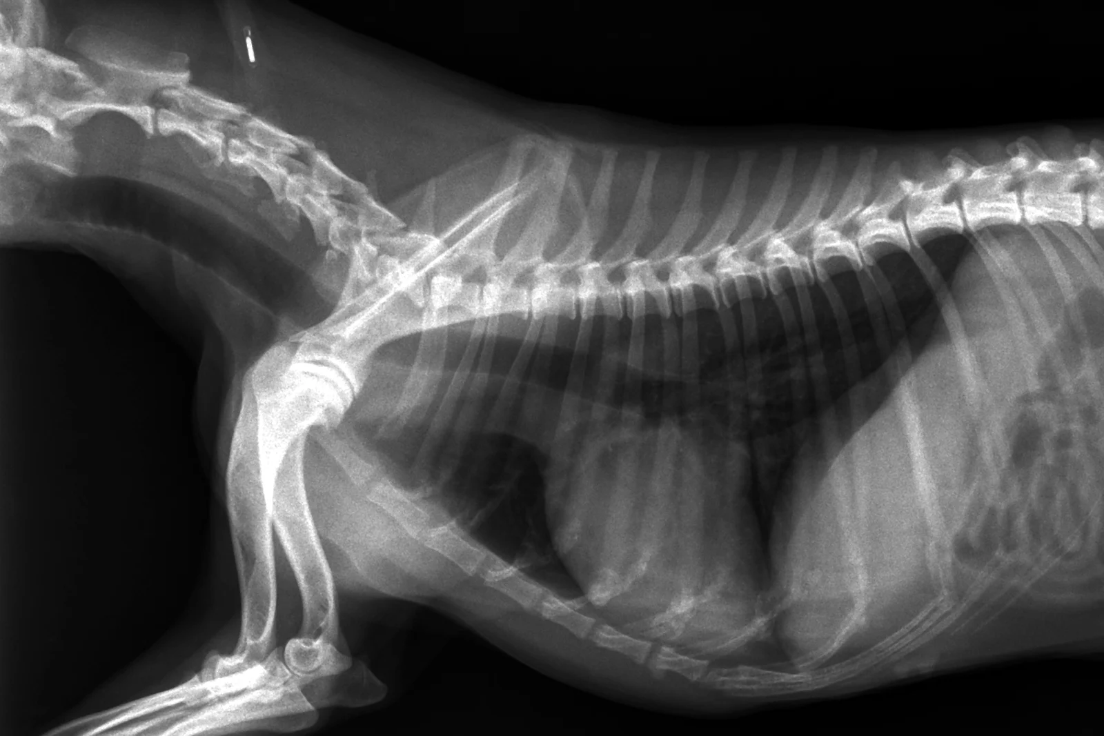
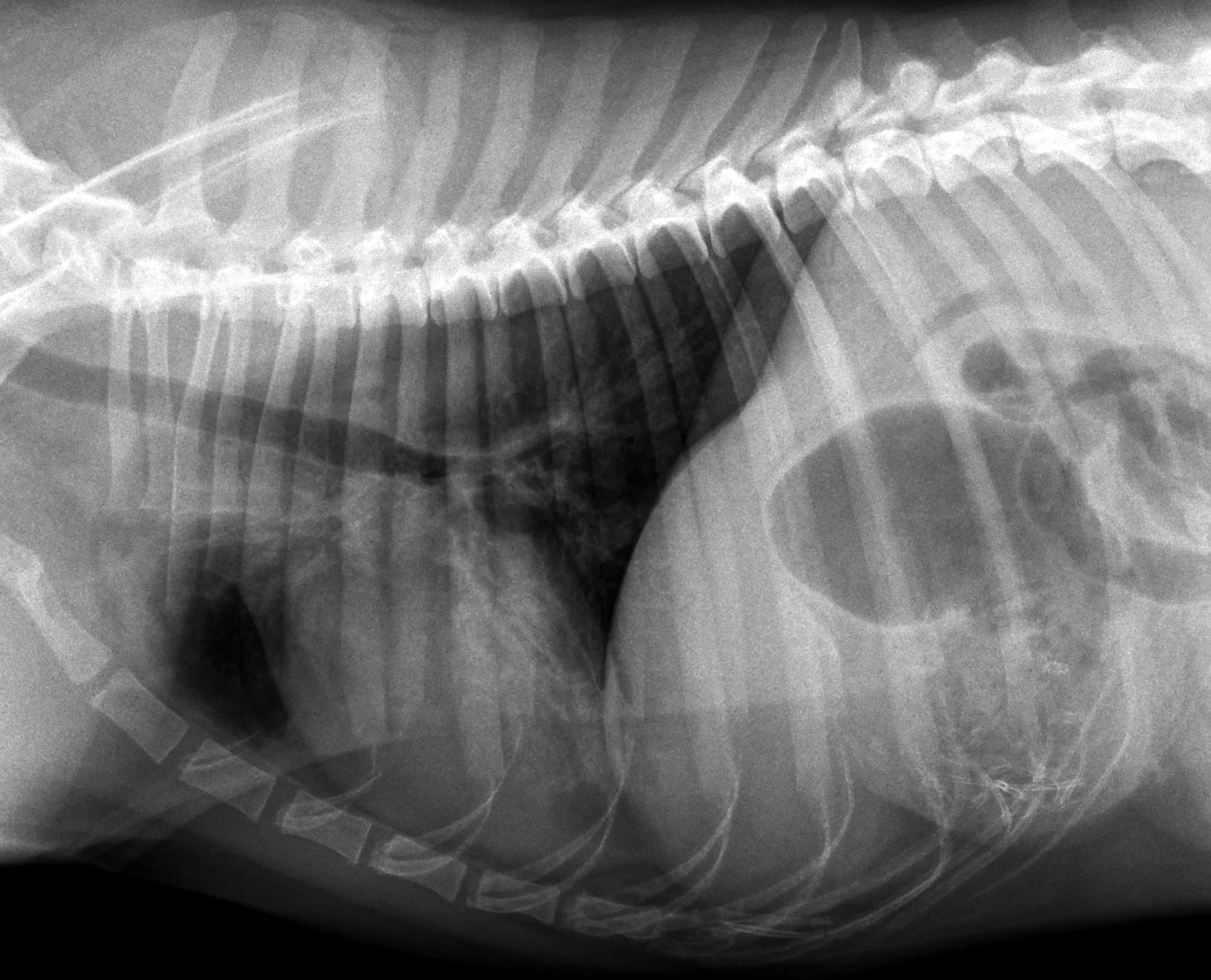
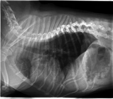
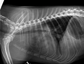
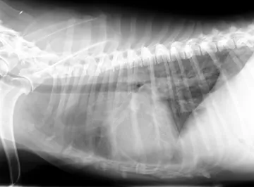
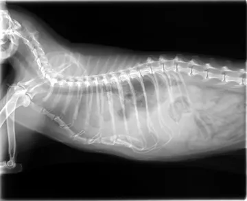
Air spaces filled. The alveoli fill with fluid or cells, so a region of lung turns to soft-tissue opacity and only the air in its bronchi remains dark.
What to look for
- Air bronchograms: dark branching bronchi against white lung.
- A lobar sign: a lobe opaque up to a sharp line at its border.
- Loss of the outline of the heart or vessels the lesion touches.
Common causes
Pneumonia (often cranioventral), cardiogenic pulmonary oedema (perihilar in dogs), haemorrhage, some neoplasia.
Against the normal
Normal lung is dark, with vessels and airways seen as fine lines and dots.
Radiographs: VetXRay, Banzato T., Burti S., Zotti A., Wodzinski M., University of Padua, CC BY 4.0. Converted to web images; findings as annotated by the dataset's radiologist. Dataset · Licence. For learning: not a diagnostic service.