Thorax
Bronchial pattern
Thickened airway walls. The walls of the bronchi are thickened or infiltrated, so airways that are normally hard to see become visible across the lung.
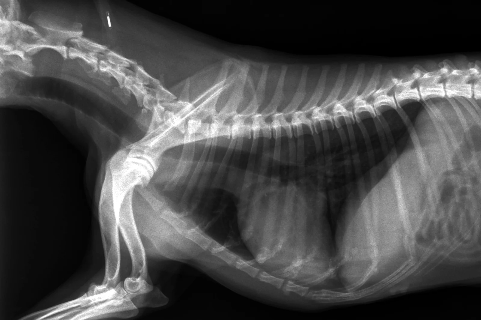
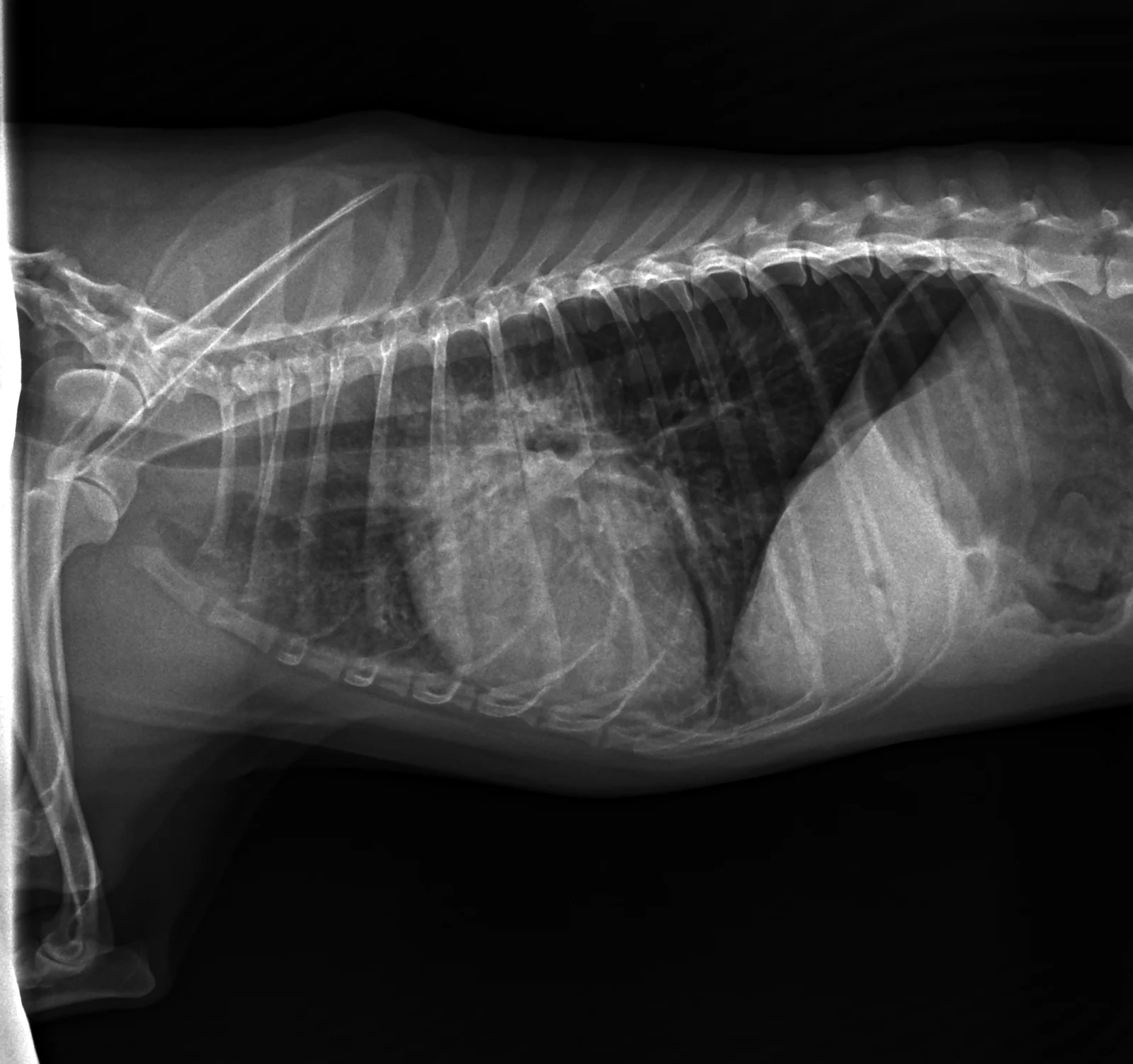
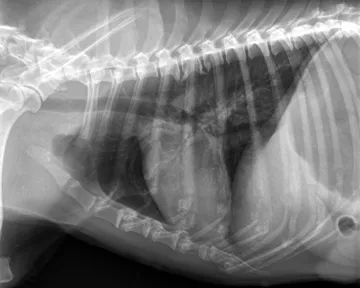
Two different patients, so size and positioning differ as well as the finding. Compare what the finding changes, not the dog.
What to look for
- Doughnuts: airways seen end on as rings.
- Tram lines: parallel lines of airways seen side on.
- More visible in the periphery of the lung than normal.
Common causes
Chronic bronchitis, feline asthma, infection, ageing changes in older dogs.
Against the normal
Normal bronchial walls are thin and seen mostly near the hilus.
Radiographs: VetXRay, Banzato T., Burti S., Zotti A., Wodzinski M., University of Padua, CC BY 4.0. Converted to web images; findings as annotated by the dataset's radiologist. Dataset · Licence. For learning: not a diagnostic service.