Thorax
Megaoesophagus
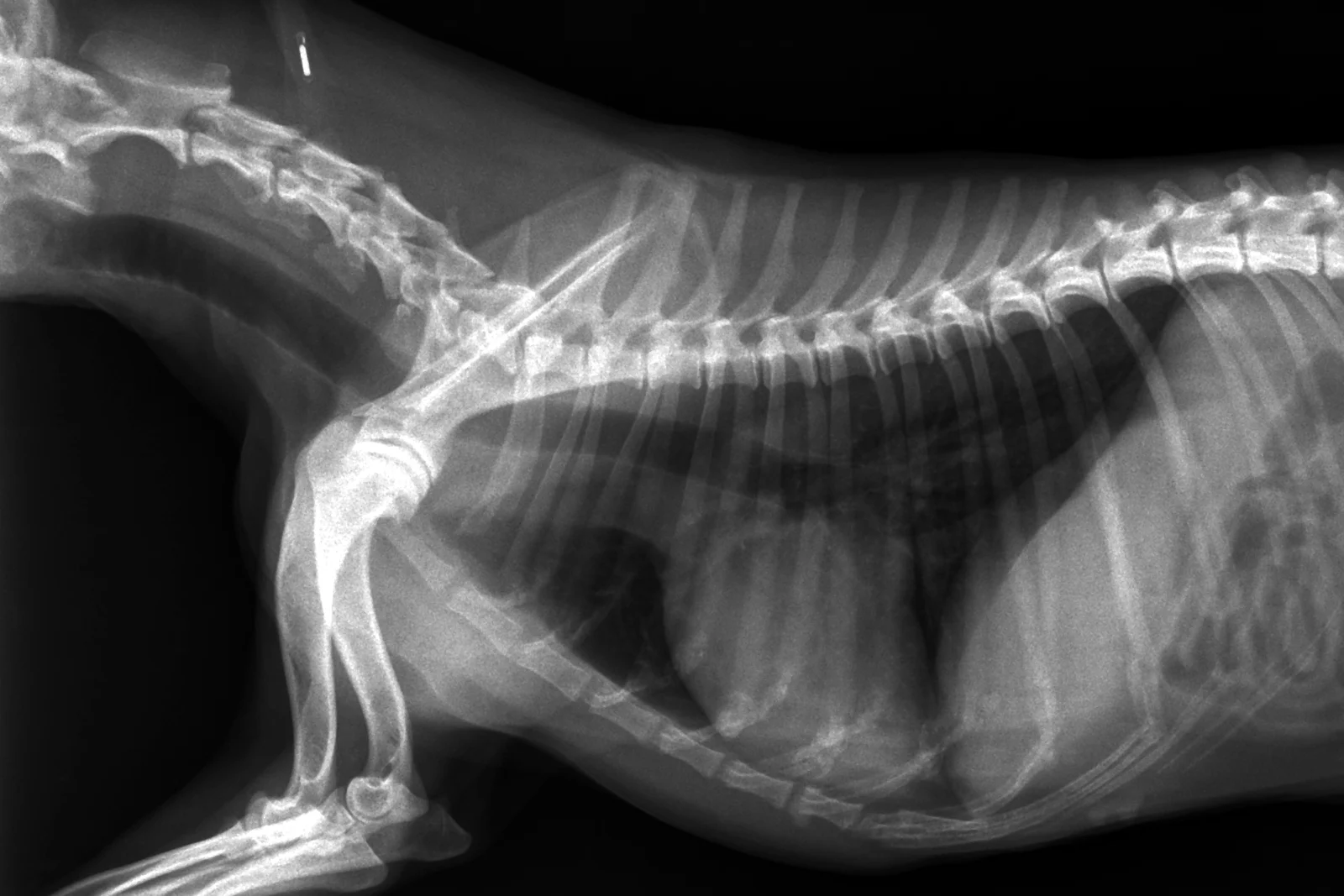
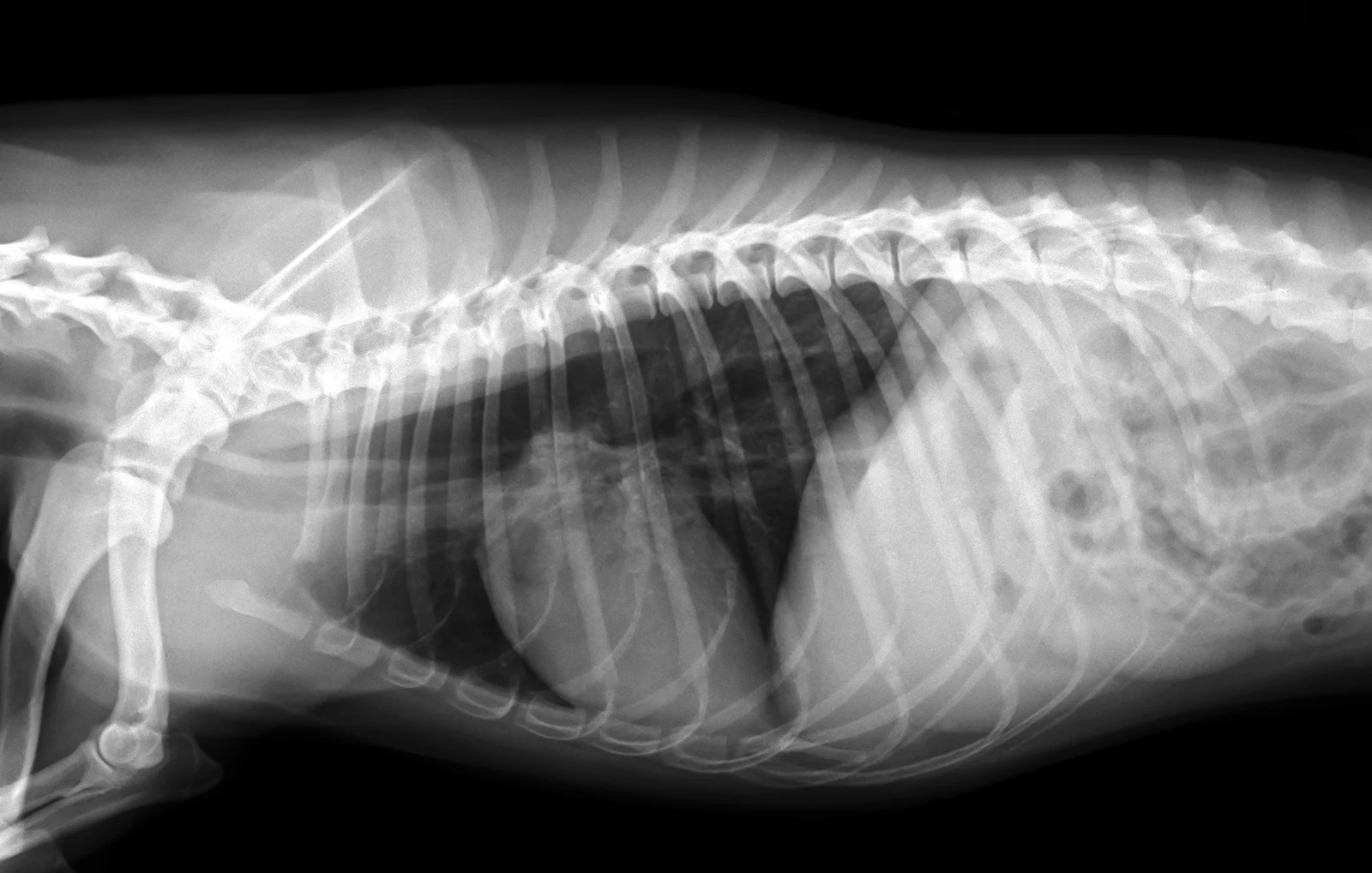
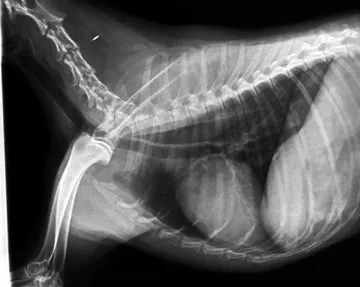
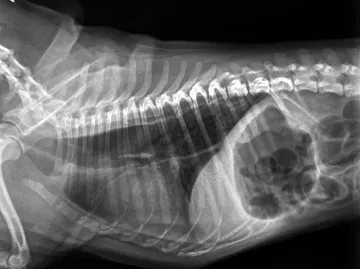
A dilated oesophagus. The oesophagus is enlarged and does not empty, so it shows on the film filled with air, fluid or food, where a normal oesophagus is barely seen.
What to look for
- A gas-filled tube dorsal to the trachea along the thorax, with visible walls.
- The tracheal stripe sign: the wall of the oesophagus and the wall of the trachea summating into one thick line.
- The trachea pushed ventrally; aspiration pneumonia in the cranioventral lung is common with it.
Common causes
Idiopathic (congenital or acquired), myasthenia gravis, hypoadrenocorticism, vascular ring anomalies in puppies.
Against the normal
A normal oesophagus is usually not visible, or only as a small amount of air.
Radiographs: VetXRay, Banzato T., Burti S., Zotti A., Wodzinski M., University of Padua, CC BY 4.0. Converted to web images; findings as annotated by the dataset's radiologist. Dataset · Licence. For learning: not a diagnostic service.