Vetomica Imaging · Ultrasound · Abdomen and heart
Ultrasound, organ by organ.
Real dog and cat scans. For each organ, learn the normal image first, then see what the common findings look like beside it.
Kidney
Read the outline, the cortex against the medulla, and the pelvis. A normal kidney is smooth and bean-shaped, with a brighter cortex around a darker medulla and almost no visible pelvis.
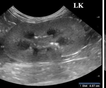 Normal
- Smooth, even outline
- Cortex brighter than the medulla, with a clear border between them
- Pelvis barely visible
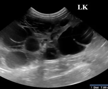 Finding
- Many round, black (anechoic) cysts of different sizes
- Cysts distort the outline and the corticomedullary border
- Usually both kidneys; Persians and related breeds carry the PKD1 variant
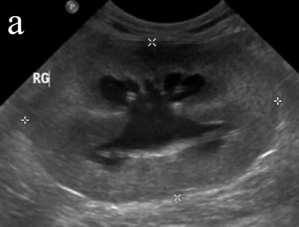 Finding
- Pelvis widened into a black, branching space
- Thinned rim of tissue around it
- Look down the ureter for the cause: a stone, a stricture or a mass
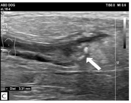 Finding
- Bright (hyperechoic) focus inside the ureter
- Clean acoustic shadow behind it
- Ureter and pelvis dilated above the stone
Urinary bladder
Judge the wall only when the bladder is well filled: a contracted bladder always looks thick. Normal urine is black, the wall thin and even.
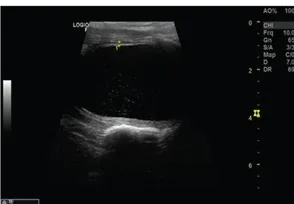 Normal
- Wall thin and regular (about 1.7 mm here)
- Urine anechoic; a little suspended sediment is common
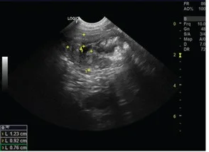 Finding
- Wall thick and irregular, worst cranioventrally in most cystitis
- Echogenic sediment on the floor or stuck to the mucosa
- Same dog as the normal image, before treatment
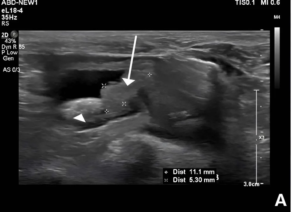 Finding
- Irregular, hyperechoic nodule rising from the wall at the trigone
- Calculi beside it (arrowhead)
- A trigonal mass in an older dog raises urothelial carcinoma until a sample says otherwise
Liver and gallbladder
Compare the liver with its neighbours: normally it is a little darker than the spleen and similar to the right kidney's cortex, with crisp portal vein walls and sharp edges.
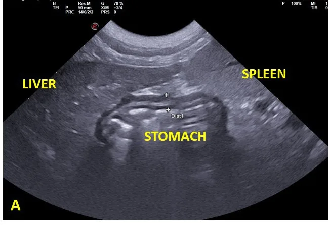 Normal
- Even, fine texture
- Less echogenic than the spleen
- Thin, tapered margins
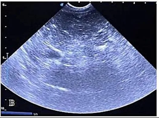 Finding
- Whole liver brighter than normal
- Portal vein walls fade into the background
- Here grade 3 lipidosis; steroid hepatopathy and other infiltrates look alike
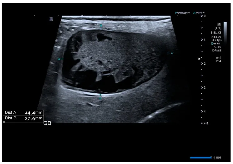 Finding
- Immobile echogenic bile with a stellate centre: the 'kiwi fruit' pattern
- Does not move when the dog is turned
- Normal bile is black and moves with gravity
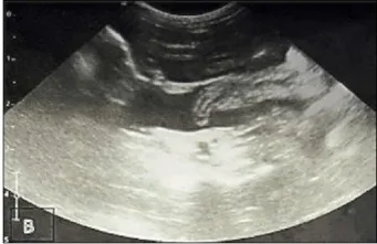 Finding
- Black fluid outlining liver lobes and bowel
- Collects in the lowest part of the abdomen
- Here with cirrhosis; sample it to tell transudate, exudate and blood apart
Spleen
The spleen is the brightest abdominal organ, with a fine, even texture and a smooth capsule. A mass in it may be benign or malignant: ultrasound alone rarely tells them apart.
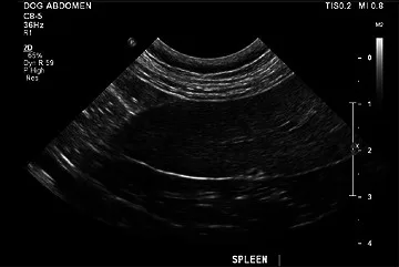 Normal
- Fine, homogeneous texture
- Brighter than the liver
- Smooth, thin capsule
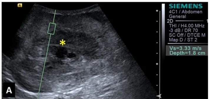 Finding
- Mass of mixed, heterogeneous echogenicity
- Cavitated, fluid-filled areas
- Check the abdomen for free fluid and the right atrium for a second mass
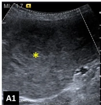 Finding
- Also mixed and heterogeneous: on B-mode it can look like haemangiosarcoma
- Elastography and contrast help; histology decides
Stomach and intestine
The gut wall has five layers, alternately bright and dark: mucosal surface, mucosa, submucosa, muscularis, serosa. Losing that layering is the most important sign of all.
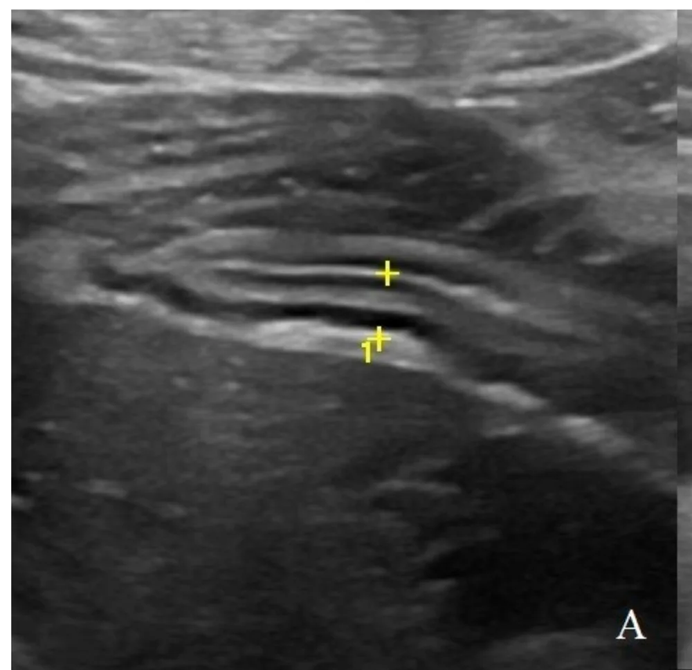 Normal
- All five layers visible
- Wall about 4 mm thick between the cursors
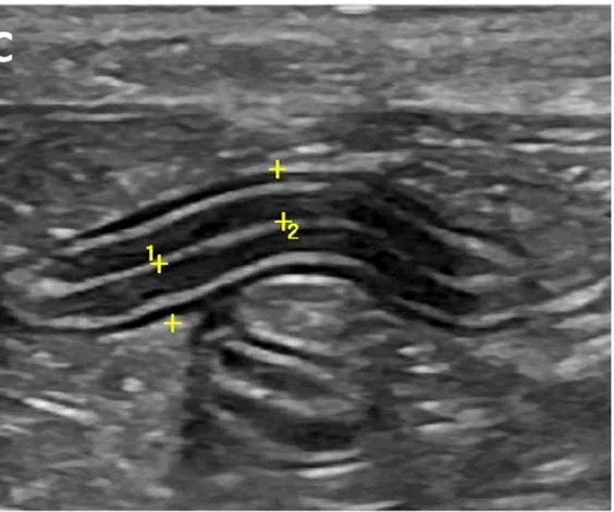 Normal
- Five layers in a thin, even wall
- Collapsed lumen, no dilation
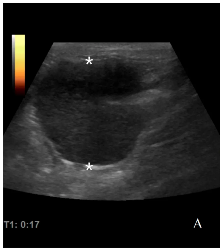 Finding
- Thick, hypoechoic wall mass (24 mm between the asterisks)
- Layering lost completely
- Lymphoma tends to be dark and uniform; carcinoma more mixed
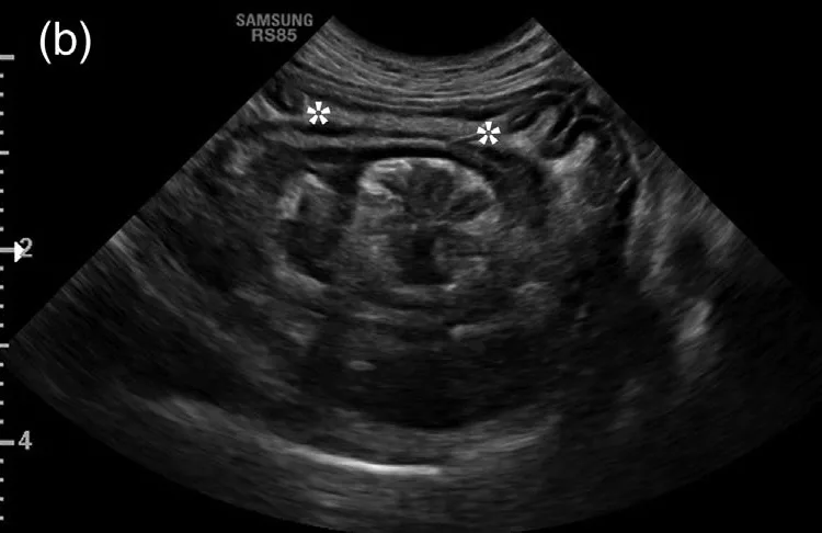 Finding
- Target of concentric rings in cross-section: one segment inside another
- Hyperechoic fat trapped between the layers (asterisks)
- Here gastrogastric; in young dogs it is usually ileocolic
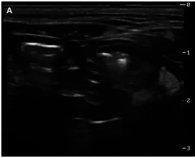 Finding
- Dilated, fluid-filled loops
- Little or no peristalsis
- Follow the loops to where calibre changes: the cause sits there
Adrenal glands
Find each adrenal beside the aorta (left) or caudal vena cava (right). A normal gland is small, peanut- or crescent-shaped and evenly dark.
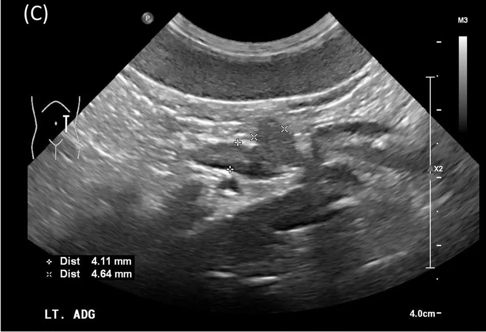 Normal
- Crescent-shaped, homogeneous and hypoechoic
- Poles about 4–5 mm thick
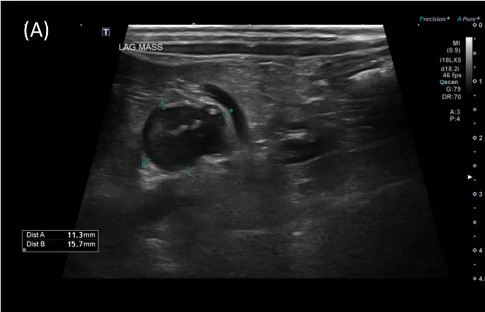 Finding
- Rounded, enlarged gland with solid and cystic parts
- Pressing on the neighbouring vessel: check it for invasion
- Here a pheochromocytoma
Heart
Echocardiography reads chamber size, wall thickness and valve flow. Start from the right parasternal long-axis view.
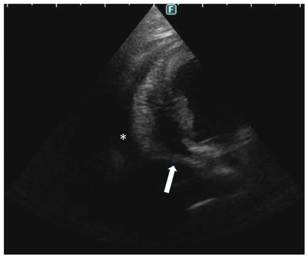 Finding
- Black band of fluid around the heart (asterisk)
- Right atrium collapsing (arrow): tamponade
- Look for a mass on the right atrium or the heart base
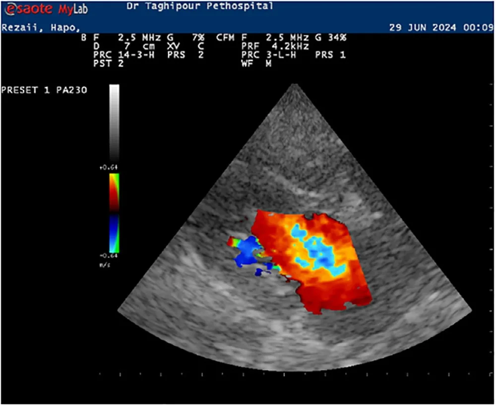 Finding
- Colour jet back into the left atrium in systole
- Thickened mitral leaflets in myxomatous valve disease
- Measure the left atrium against the aorta (LA:Ao)
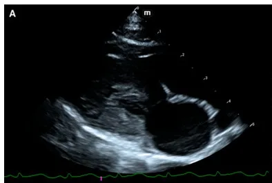 Finding
- Thick left ventricular walls, a small cavity
- Enlarged left atrium
- Over 6 mm diastolic wall thickness is the usual cut-off in cats
Every image is a panel cropped from a figure in an open-access paper, used under CC BY 4.0; the paper is linked under each image. Labels and arrows are the authors'. For learning: not a diagnostic service.
Sources: Jaturanratsamee K. et al., Vet World 2026;19(4):1495–503; Pulido Vega D. et al., Front Vet Sci 2025;12:1518713; Park C. et al., Vet Sci 2026;13(1):69; Soares L. V. et al., Case Rep Vet Med 2026:7317611; Lee S. H. et al., Vet Sci 2026;13(5):472; Gomes F. G. E. et al., Braz J Vet Med 2026;48:e005026; Thi Thao T. et al., Vet World 2026;19(6):2479–95; Choe J. M. et al., Animals 2025;16(1):31; Turner R. B. S. et al., Vet Radiol Ultrasound 2026;67(4):e70202; Ercolin A. C. M. et al., Animals 2024;14(2):312; Simeoni F. et al., Animals 2021;11(3):670; Esteves-Monteiro M. et al., Vet Q 2026;46(1):2622732; Huguet E. E. et al., Vet Med Sci 2022;8(6):2251–5; Kang M. H. et al., Front Vet Sci 2024;11:1358797; Chae Y. et al., Ir Vet J 2025;78:32; Yoshida T. et al., Vet Sci 2026;13(7):722; Elyasi B., Vet Med Sci 2026;12(4):e70922; Sussadee M. et al., Animals 2026;16(12):1815.