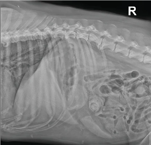
Peritoneal effusion
Findings are Pro only. Every normal study is free; Pro opens each finding beside its normal.
See ProWhat to look for
- Serosal detail lost in the ventral abdomen: fluid hides the organs' edges
- Here from a ruptured gallbladder (bile peritonitis)
- Only a sample says what the fluid is; ultrasound finds small amounts
Against the normal
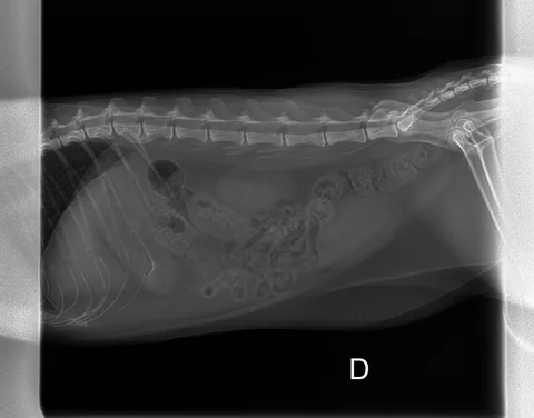
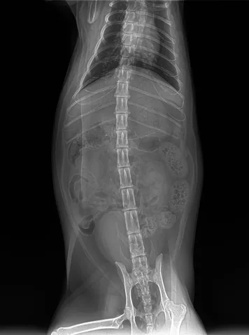
Read serosal detail first: fat lets you see the organs' edges, and losing it means fluid, no fat, or a crowd. Then size and place each organ against the vertebrae and the last rib, and follow the gas through the gut.
Source: Vekšins A. et al., Case Rep Vet Med 2025. CC BY 4.0. For learning: not a diagnostic service.
Read next
Read the film, then see the anatomy in 3D.
Vetomica puts every abdomen structure on this x-ray in a complete 3D dog you can peel, cut and X-ray from any angle, with every imaging finding beside its normal and practice at reading films.
The 3D animals are free for 7 days, no card. Imaging findings are Pro; every normal study stays free.