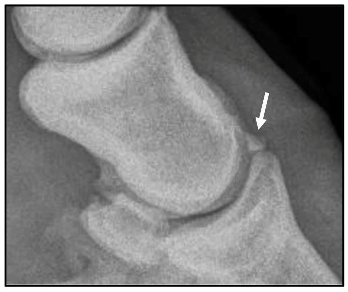
Extensor process fragment
Findings are Pro only. Every normal study is free; Pro opens each finding beside its normal.
See ProWhat to look for
- A 5 mm bone fragment at the extensor process of the distal phalanx (arrow)
- Inside the distal interphalangeal (coffin) joint
- Here removed by arthroscopy
Against the normal
Read every bone's outline and density, then every joint: the space, the margins and the bone under the cartilage. Compare with the other limb, and always take at least two views at right angles.
Source: Phillips N. A. I. et al., Animals 2026;16(8):1168. CC BY 4.0. For learning: not a diagnostic service.
Read next
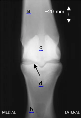 X-rayHorse · Bones and jointsNormal fetlockNormal
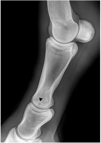 X-rayProHorse · Bones and jointsSubchondral bone cystFinding
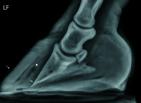 X-rayProHorse · Bones and jointsLaminitis: pedal bone rotationFinding
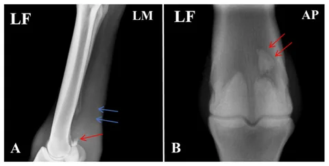 X-rayProHorse · Bones and jointsProximal sesamoid fractureFinding
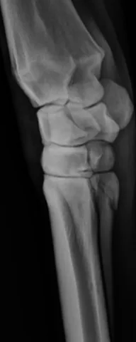 X-rayProHorse · Bones and jointsSplint bone fractureFinding
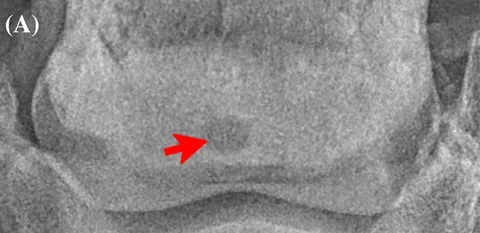 X-rayProHorse · Bones and jointsNavicular bone lesionFinding
Read the film, then see the anatomy in 3D.
Vetomica puts every bones and joints structure on this x-ray in a complete 3D horse you can peel, cut and X-ray from any angle, with every imaging finding beside its normal and practice at reading films.
The 3D animals are free for 7 days, no card. Imaging findings are Pro; every normal study stays free.