Chest
Chest films, and the heart on ultrasound. Start from the normal, then each problem beside it.
Normal canine thorax, lateral view
Choose a number to name the structure.
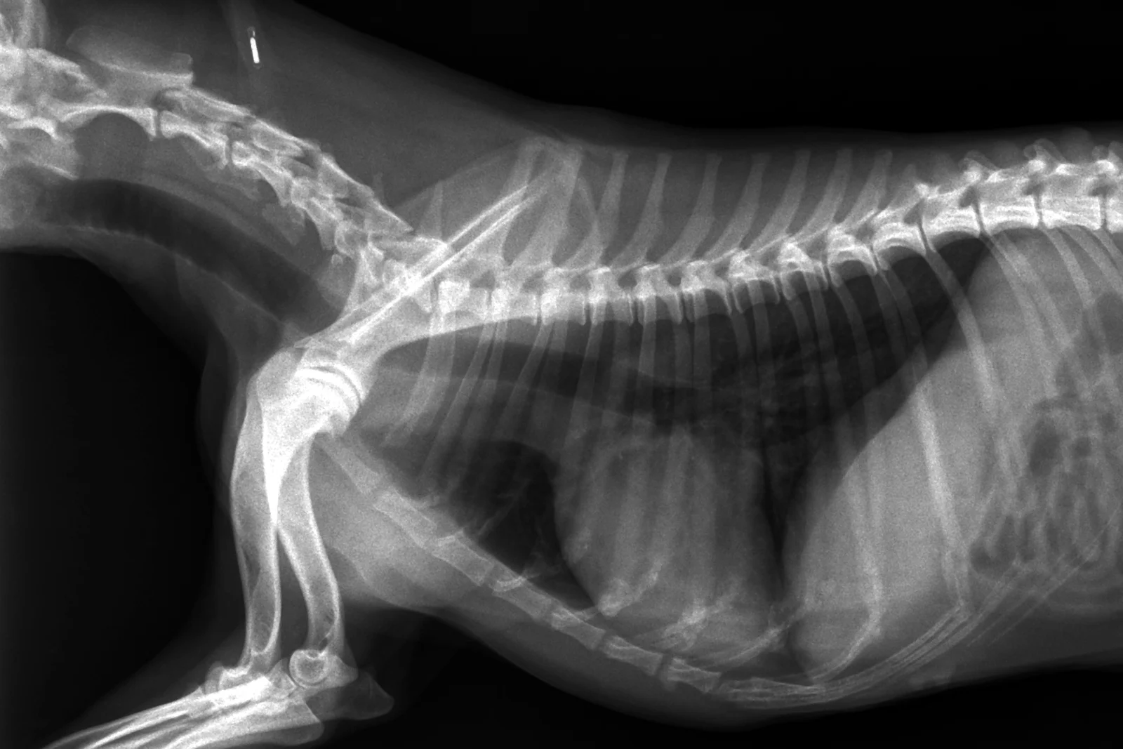
Heart Cor
The cardiac silhouette: about two and a half to three and a half intercostal spaces wide on a lateral view.
See it in 3DNormal
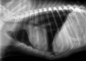 X-rayDog · ChestNormal chest, lateral (1 of 3)Normal
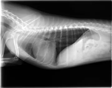 X-rayDog · ChestNormal chest, lateral (2 of 3)Normal
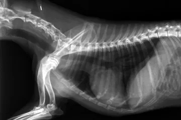 X-rayDog · ChestNormal chest, lateral (3 of 3)Normal
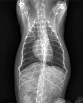 X-rayDog · ChestNormal chest, dorsoventralNormal
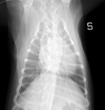 X-rayDog · ChestNormal chest, ventrodorsalNormal
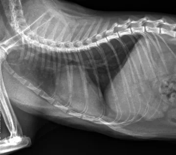 X-rayCat · ChestNormal chest, lateral (1 of 2)Normal
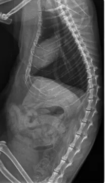 X-rayCat · ChestNormal chest, lateral (2 of 2)Normal
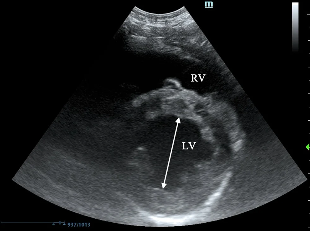 UltrasoundDog · ChestNormal left ventricleNormal
Problems
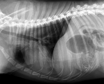 Alveolar patternAir spaces filledX-ray
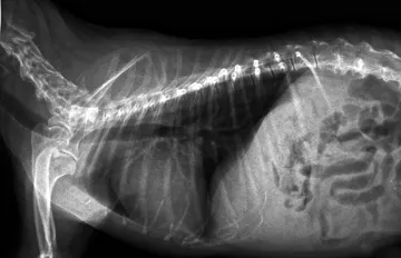 CardiomegalyAn enlarged heartX-ray
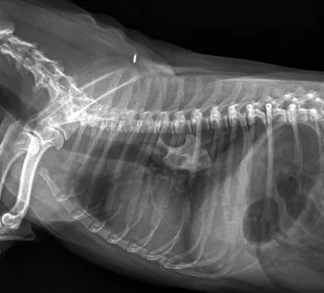 Foreign bodySomething that should not be thereX-ray
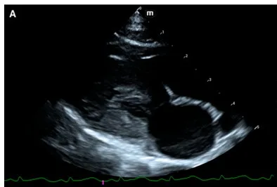 Hypertrophic cardiomyopathyThickening of the left ventricular wall, the commonest heart disease of cats.Ultrasound
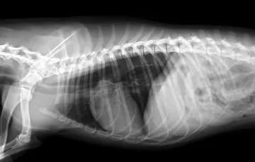 MegaoesophagusA dilated oesophagusX-ray
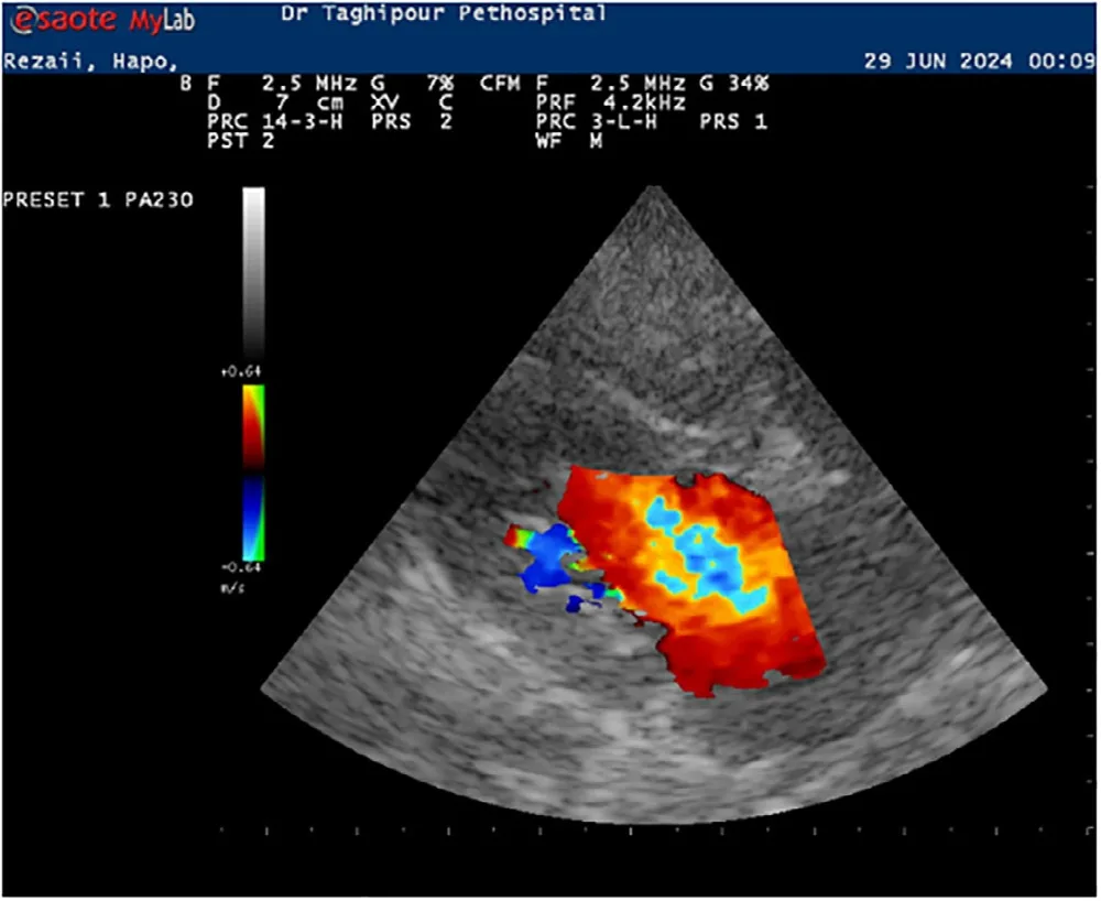 Mitral regurgitationBlood leaking back through the mitral valve into the left atrium, most often from myxomatous valve disease.Ultrasound
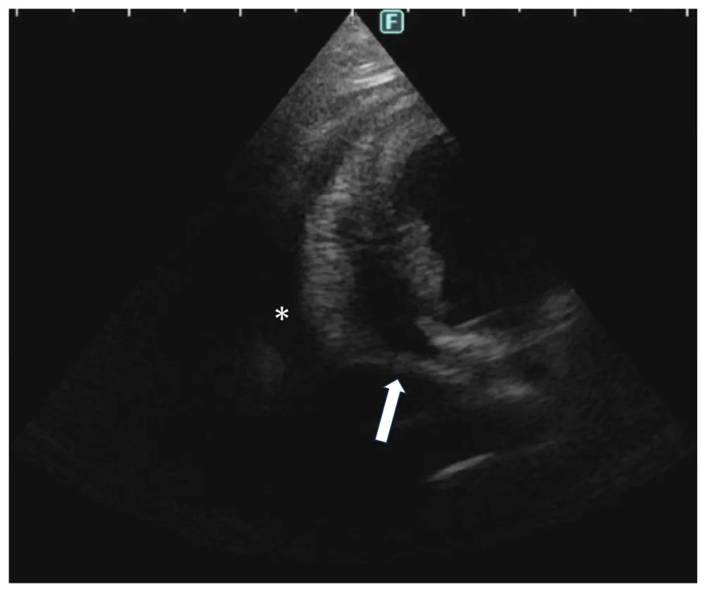 Pericardial effusionFluid in the sac around the heart.Ultrasound
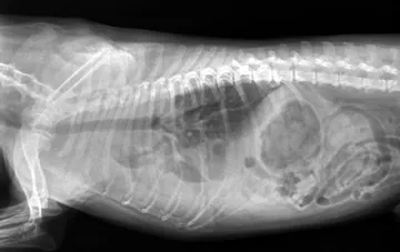 Pleural effusionFluid in the chestX-ray
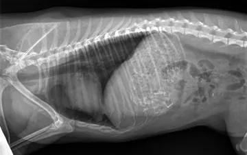 PneumothoraxAir around the lungsX-ray
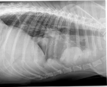 Thoracic massA mass in the chestX-ray